


By Mili Jayadeep | Science Editor
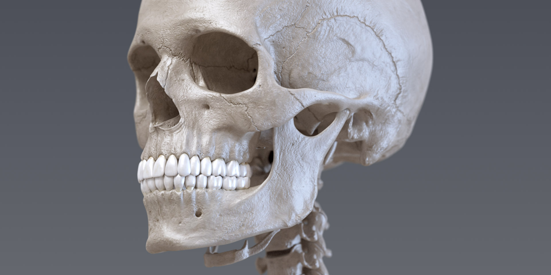
Bones of the skeleton contribute to the mechanical functioning of the body and facilitate everyday activities due to its ability to undertake loading and mechanical stress and strain owing to its structural properties. Cells within the bone have functions that result in the production and the maintenance of healthy bone. These cells include osteoblasts, osteoclasts and osteocytes, all of which have differing properties to maintain an optimum homeostasis of bone production and resorption. However, when this balance is disrupted due to pathology, such as that observed in osteoporosis, it can result in low bone mass and low bone mineral density causing weak and brittle bones. The structural changes resulting from osteoporosis affects the mechanical properties of the bone resulting in an increased risk of fracture. In the UK, over 500,000 people require treatment following a fragility fracture due to osteoporosis each year. Current research treatments are limited to preventing the progression of the condition hence research in this area is crucial. 
A team of researchers at the University of Massachusetts AmherstÔÇÖs Institute for Applied Life Sciences (IALS). This research published in the journal Science Advances could contribute to the development of therapeutics in osteoporosis.
They developed a tissue engineered model that mimics the remodeling processes that are present within healthy bone tissue. This model is a trabecular bone organoid model created to reside on osteoporotic bone. Researcher, Jungwoo Lee explains:
“Bone is a multifunctional tissue not only maintaining mechanical stability, but also regulating blood-forming and blood mineral content. However, investigating bone-remodeling biology is challenging because this process occurs inside the bone cavity. Hard and opaque bone tissue is difficult to access, thus creating realistic bone tissue models outside of the body will advance our understanding of fundamental bone biology, as well as provide new opportunities to model disease progression and screening drug responses.”
The team used bovine bone samples and demineralized it. Using thin sections of this bone, they were able to create a novel biomaterial that is closest in resemblance to the complex structural matrix of bone tissue. This material is durable, semi-transparent, enabling easy monitoring of cell processes on the model that is also of a specific thickness and surface area. The purpose of this model was to replicate the collagen matrix in unmineralized bone. This model helps to facilitate the study of bone by creating a very specific environment upon which bony cells can reside. Due to the intrinsic complexities of the environment within bone tissue, it is often challenging to study the cells that make up bone. Therefore, the provision of such a model can help study the processes within these cells, what occurs during bone remodeling and how this can be applied to develop drug targets for bone diseases such as osteoporosis.
Although the scientists have created a novel biomaterial that greatly contributes to current research into bone biology, further research is needed on human bone models to translate their work to be able to apply this to the human condition. The successful development of their research could have positive implications for developing drugs for osteoporosis as it could help reduce the screening period required for developing these therapeutics. 


Add Comment